

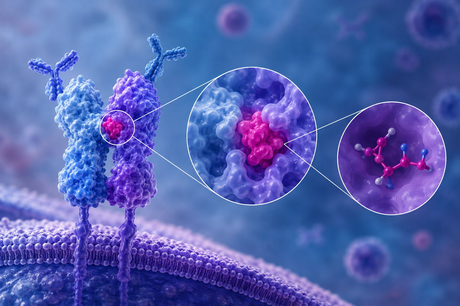
Despite advances in our understanding of the mechanisms of chronic antibody-mediated rejection due to the development of Human Leukocyte Antigen (HLA) alloantibodies, it remains a leading cause of solid-organ late-stage graft loss1. Within the first 10 years after transplantation, studies have shown that up to 30% of previously non-sensitised kidney transplant recipients develop de novo donor-specific antibodies (DSA). Around 40% of these then go on to lose their grafts within 5 years of developing that DSA2. The most frequently observed de novo DSA produced after transplantation has been found to be those towards HLA-DQ2,3,4. Not only are they the predominant de novo DSA, but they have also been associated with higher frequency of pathogenicity and graft loss as compared to DSA directed towards other HLA loci. The reasons for this are not yet fully understood, but structural differences, differential expression levels and distinct intracellular signalling pathways induced by T-cells and alloantibody interactions are all under investigation5,6. It is also worth noting that whilst HLA-DQB1 is the most emphasised target of HLA-DQ antibodies, HLA-DQA1 antibodies are independently associated with increased risk of antibody-mediated rejection in their own right7,8.
HLA epitopes and eplets
In solid-organ transplantation, low-resolution matching at HLA-A, -B & -DRB1 is often still the norm as donor allocation systems were historically set up based on HLA typing techniques which classified HLA under broad serological groupings. However, due to advancing technologies the availability of high-resolution donor and recipient HLA typing across the 11 key loci, including HLA-DQ, is now commonplace; enabling consideration of HLA matching in significantly more depth. For example, Rene Duquesnoy et al. developed the HLA Matchmaker algorithm using in silico techniques to describe the clusters of polymorphic amino acids (~15 angstroms diameter) which distinct HLA molecules consist of, termed epitopes. Where they differ between donor and recipient, these epitopes represent the full footprint recognised by an alloantibody. Each ‘structural epitope’ can be further refined to include a smaller footprint termed the ‘eplet’ (~3 angstroms diameter), sometimes referred to as the ‘functional epitope’, which has been thought to be particularly crucial for determining alloantibody specificity and binding strength9,10.
HLA-DQ matching and alloantibody responses
Eplet mismatches in HLA-DQ have been found to be associated with increased risk for de novo DSA formation, graft rejection and graft failure after kidney transplantation. This association has been seen most strongly with HLA-DQ eplet mismatches as compared to other loci and the potential utility of considering HLA-DQ eplet mismatch load in personalised post-transplant immunosuppression strategies has been highlighted11. The discovery of new HLA-DQ eplets is still very much ongoing. HLA eplets are described in the HLA Eplet Registry database after allele-specific amino acid sequences are aligned to highlight the surface accessible polymorphic residues. The database, however, is not exhaustive and different methods of identifying eplets are required for those where the peptide itself has been shown to be involved alongside the HLA molecule and where the eplet spans the alpha- and beta-chains. In addition, not all eplets within the database have been experimentally verified i.e. antibody verified12,13,14. Whilst a comprehensive database of HLA eplets is critical for predicting and interpreting HLA antibody responses, an appreciation of their relative, and evidently unequal, ability to induce an antibody response i.e. their immunogenicity is also crucial. Tambur et al. posit that identifying a ‘pro’ group of eplets empirically proven to be associated with induction of an antibody response when mismatched is a more clinically useful method of risk stratification than purely enumerating mismatch load. Their work also demonstrates that secondary to an initial inflammatory response whereby a DSA response is produced, a secondary antibody response can be formed against the same donor allele in a process termed epitope spreading. They hypothesise that generation of the initial DSA response to a ‘pro’ group eplet induces a more inflammatory environment, creating more favourable conditions for a secondary antibody response towards less intrinsically immunogenic eplets. Through their work, the group has highlighted DQA1*05 heterodimer associated mismatched eplets as potentially highly immunogenic15,16,17.
Extended HLA-DQ panel: LABScreen™ Single Antigen HLA Class II ExPlex – Group 2
Current literature is, therefore, highlighting the importance of careful evaluation of HLA-DQ DSA responses in patients at both an allelic and eplet-level to both inform post-transplant management and characterise profiles in anticipation of re-transplantation where graft loss has occurred. LABScreen™ Single Antigen HLA Class II ExPlex – Group 2, an expanded HLA-DQ allele panel, has been developed with this in mind. Figure 1 details the HLA Class II ExPlex – Group 2 bead panel composition, highlighting where the LABScreen™ Single Antigen HLA Class II ExPlex and LABScreen™ Single Antigen Class II – Supplement panels differ and overlap.
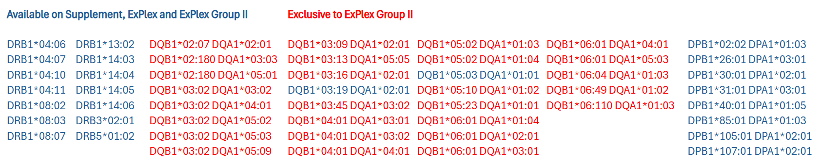
Figure 1: LABScreen™ Single Antigen HLA Class II ExPlex – Group 2 bead panel – overlaps and differences with LABScreen™ Single Antigen HLA Class II ExPlex and LABScreen™ Single Antigen Class II – Supplement.
The Group 2 panel looks to address two areas; increasing alpha-beta heterodimer diversity and the coverage of unique polymorphic positions.
Alpha-beta heterodimer diversity
LABScreen™ Single Antigen HLA Class II ExPlex – Group 2 expands the alpha-beta heterodimer diversity of the standard HLA Class II Single Antigen and ExPlex bead panels. It enables better differentiation between alpha- and beta-chain antibody reactivity through separation of overlapping bead reactivity patterns and clarification of chain-specific eplet targets of reactivity.
Figure 2 shows HLA-DQ1 antibody reactivity detected using the standard LABScreen™ Single Antigen HLA Class II panel. All DQ5 and DQ6 beads are positive (highlighted in blue). However, it is not possible to rule out whether there is antibody, alternatively to or in addition to, directed towards DQA1*01 as these are present exclusively in combination with HLA-DQ5 and -DQ6 beta chains on this panel.
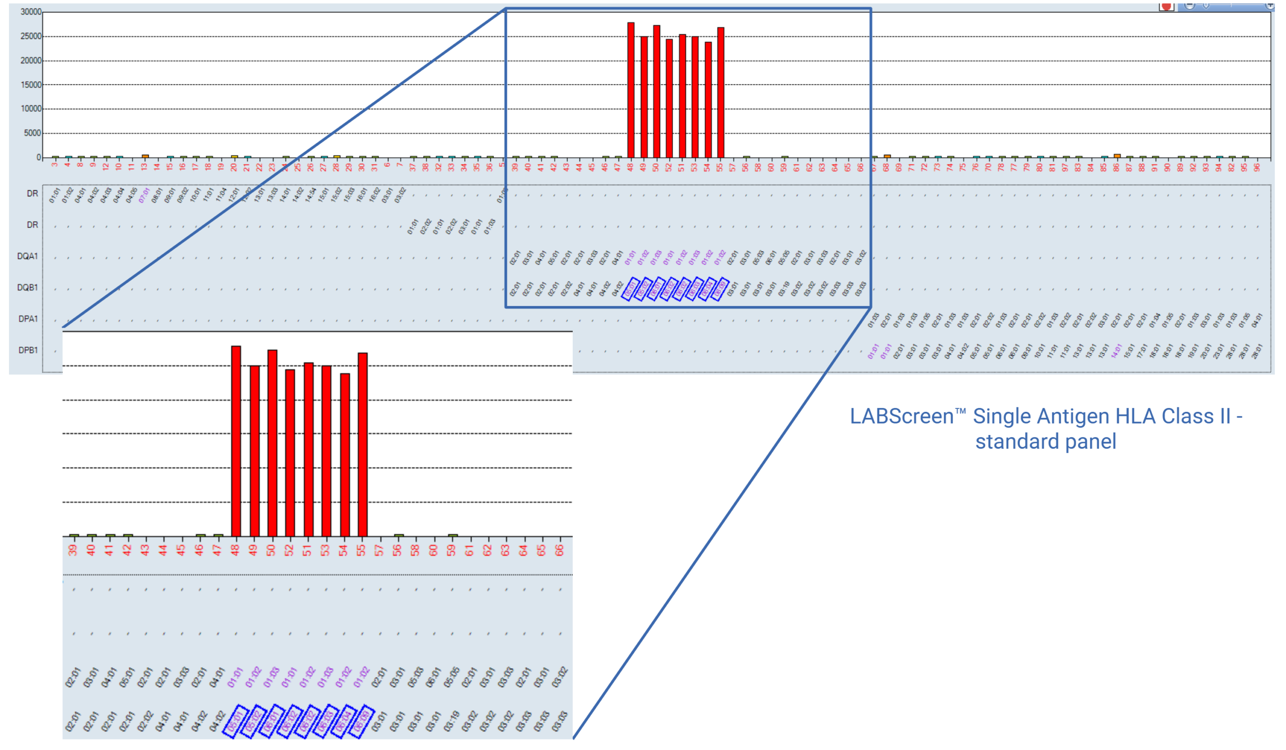
Figure 2: LABScreen™ Single Antigen HLA Class II standard panel showing DQ1 (highlighted) and potentially DQA*01 antibody reactivity.
Using the HLA Fusion™ software version 4.7.x ‘Epitope Panel’ functionality, two eplets can be easily identified which could account for the full profile of reactivity: 52PQ (beta-chain eplet found on DQ5 and DQ6) and 52SK (alpha-chain eplet found on all DQA1*01 alleles). Both eplets demonstrate identical patterns of reactivity and either or both could be responsible (Figure 3).
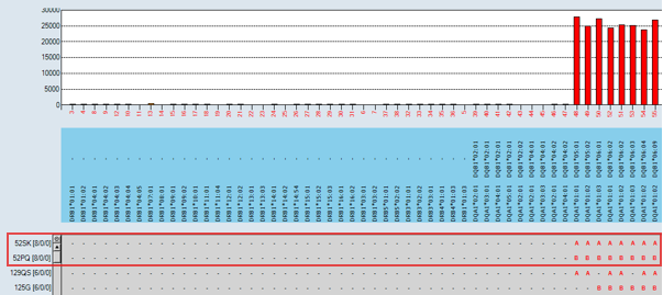
Figure 3: HLA Fusion™ 4.7.x Epitope Panel showing eplets potentially responsible for full antibody pattern of reactivity: 52PQ (beta-chain eplet found on DQ5 and DQ6) and 52SK (alpha-chain eplet found on DQA1*01).
When the same serum is tested using the HLA Class II ExPlex – Group 2 panel in combination with the standard panel, the results are as seen in Figure 4. The inclusion of DQ6 beads in combination with alternative DQA chains in the Group 2 panel rules out DQB1*06:01 as a potential target.
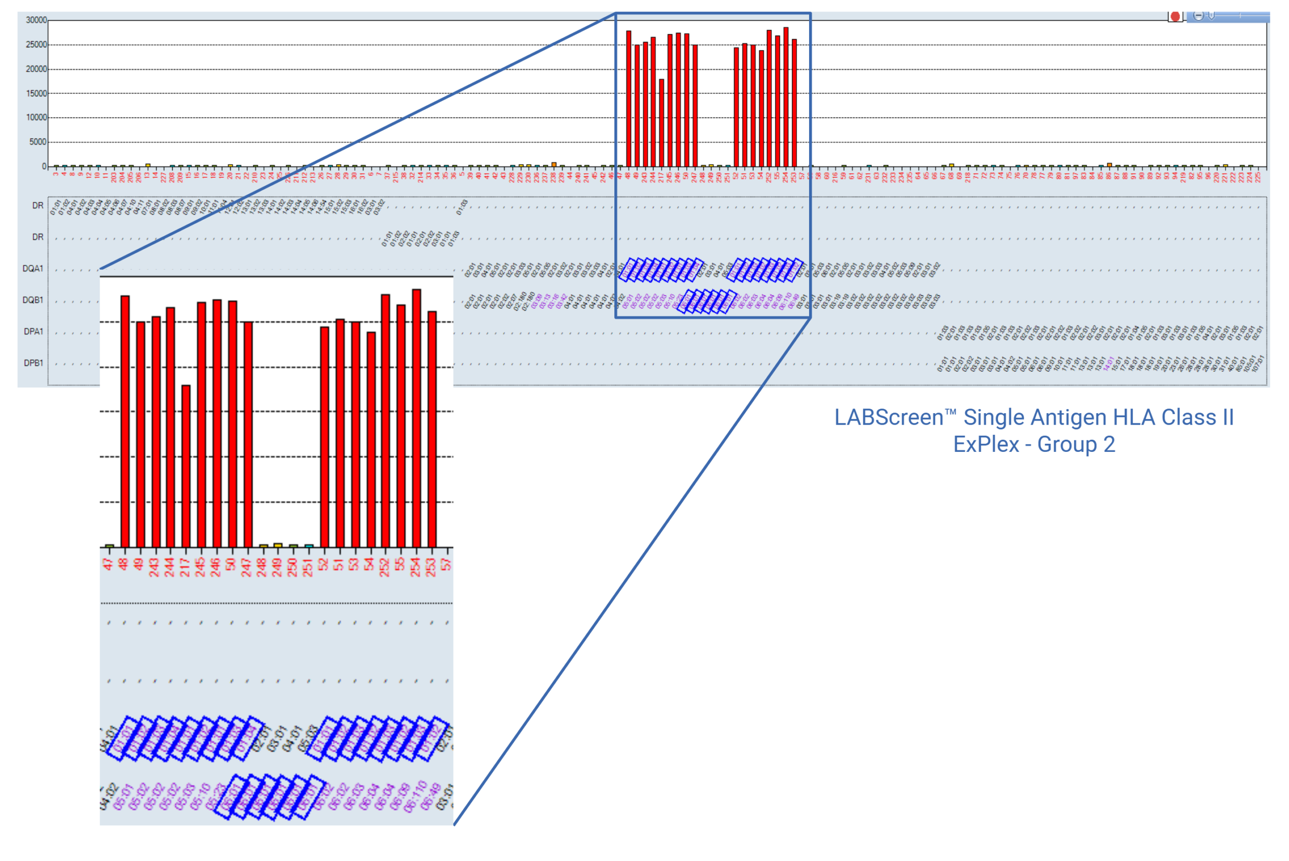
Figure 4: DQ1 broad group reactivity with LABScreen™ Single Antigen HLA Class II ExPlex – Group 2 in combination with the standard HLA Class II panel. DQB1*06:01 and DQA1*01 bead specificities highlighted.
Utilising the ‘Epitope Panel’ function again, 52PQ (beta-chain eplet) can now be ruled out based on absence of reactivity to multiple DQB1*06:01 beads (Figure 5). 52SK (alpha-chain eplet), however, remains only on positive beads including DQB1*06:01 beads in combination with DQA1*01. The addition of the HLA Class II ExPlex – Group 2 panel has, therefore, confirmed reactivity to the alpha-chain (DQA1*01) and ruled out the beta-chain potential target of reactivity (52PQ) identified by the standard panel, reducing the number of eplet ambiguities.
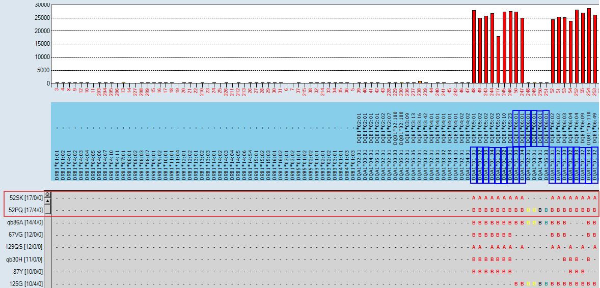
Figure 5: HLA Fusion™ 4.7.x Epitope Panel including HLA Class II ExPlex – Group 2 data. DQB1*06:01 and DQA1*01 bead specificities highlighted.
Unique polymorphic amino acid position coverage
The introduction of new alleles into the LABScreen™ Single Antigen HLA Class II ExPlex – Group 2 panel has been carefully thought through with respect to their ability to interrogate unique polymorphic amino acid positions, as opposed to considering individual allele population frequencies which has been the previous strategy for inclusion. The new specificities help to distinguish eplet patterns of reactivity by introducing alleles which, when positive or negative, are informative to the overall pattern of reactivity.
Figure 6 shows a commonly seen HLA-DQ3 antibody pattern of reactivity on the standard LABScreen™ Single Antigen HLA Class II panel. Due to the alpha-beta heterodimer combinations present on the panel, it is not possible to rule out additional reactivity to DQA1*03:02, 05:03, 05:05 or 06:01.
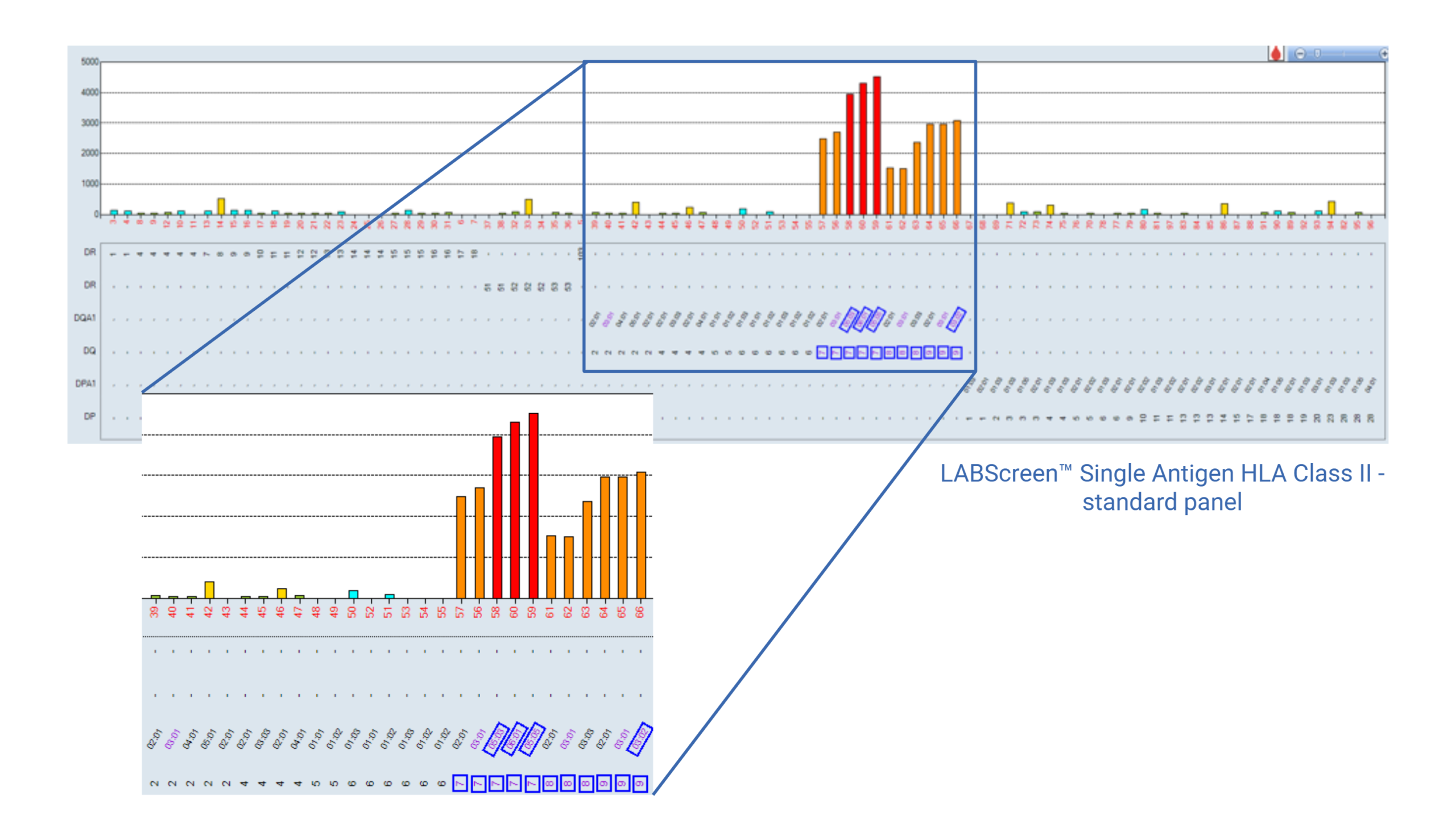
Figure 6: LABScreen™ Single Antigen HLA Class II standard panel data showing DQ3 broad group and potential DQA1*03:02, 05:03, 05:05 or 06:01 reactivity (all highlighted).
Viewing the Epitope Panel as in Figure 7, the 55PP residues are a clear potential target of reactivity. In addition, 45EV cannot be ruled out.
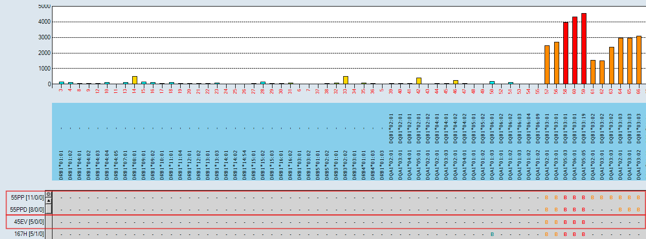
Figure 7: HLA Fusion™ 4.7.x Epitope Panel with LABScreen™ Single Antigen HLA Class II standard panel data – 55PP and 45EV identified as potential targets of reactivity.
Figure 8 shows the addition of the HLA Class II ExPlex – Group 2 panel, which contains four newly represented DQ3 broad group alleles. In this case, one of these (DQB1*03:16) is clearly negative in comparison to all other DQ3 broad group antigens. Due to the increased alpha-beta heterodimer diversity within the newly added DQ3 alleles, it is now possible to rule out DQA1*03:02 and DQA1*05:03 reactivity.
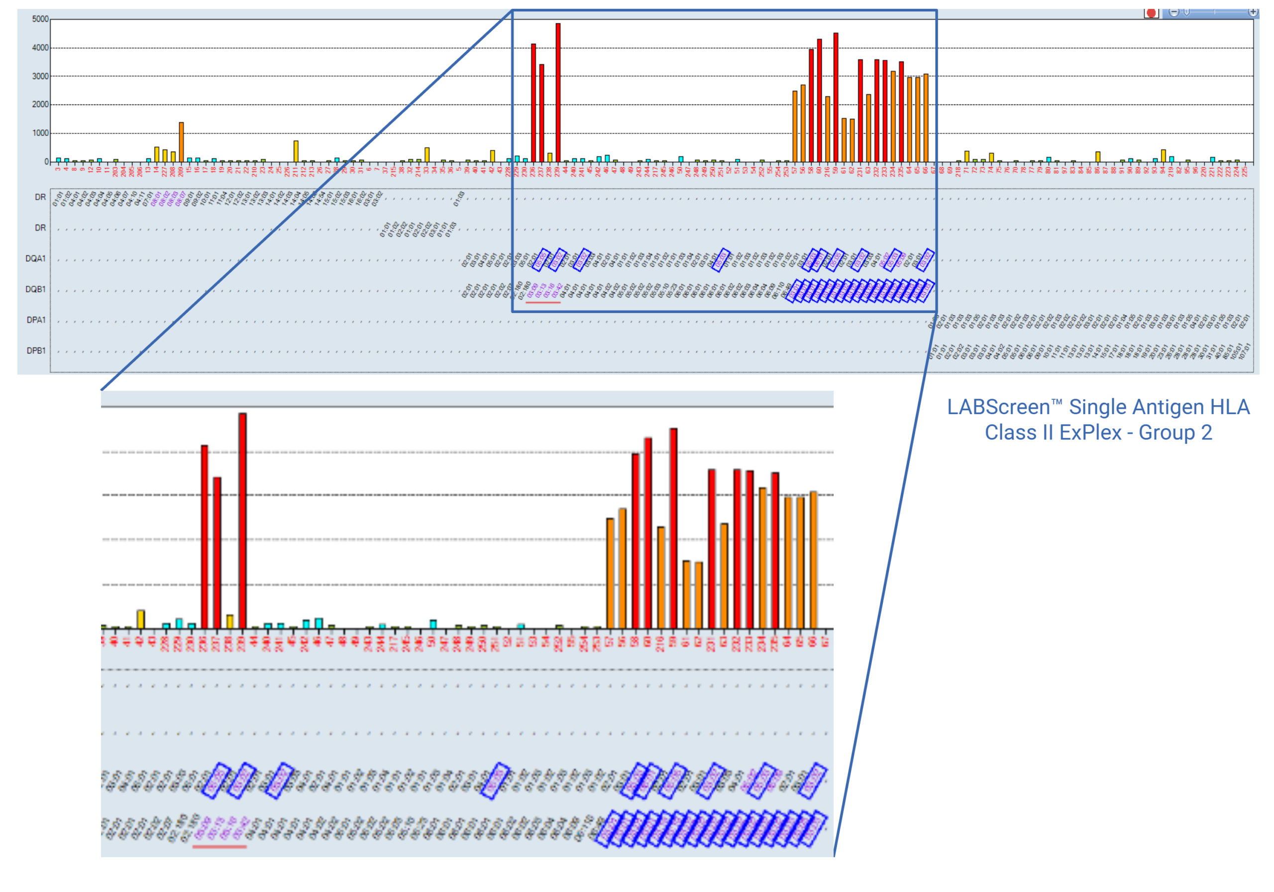
Figure 8: Partial DQ3 broad group reactivity with LABScreen™ Single Antigen HLA Class II ExPlex – Group 2 in combination with the standard HLA Class II panel. DQ3 broad group plus DQA1*03:02, 05:03, 05:05 and 06:01 specificities are highlighted or underlined.
Again, utilising the Epitope Panel (Figure 9), the lack of reactivity to DQB1*03:16 which has a glutamic acid (E) at position 45 enables 45EV to be ruled out as a target of reactivity, thereby reducing target epitope ambiguity.
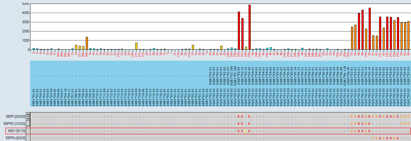
Figure 9: HLA Fusion™ 4.7.x Epitope Panel including HLA Class II ExPlex – Group 2 data: 45EV ruled out.
Whilst DQB1*03:16 is not common in all populations, this serum demonstrates the widespread utility of its addition to the panel with respect to added resolution in epitope analysis. Figure 10 shows its unique polymorphism at position 55 as compared to the other DQ3 broadoli oli group antigens on the panel i.e. a glutamine (Q) rather than a proline (P). In addition to ruling out 45EV, the polymorphism at position 55 combined with the lack of bead positivity, adds further weight to 55PP being a responsible epitope for the pattern of reactivity in this serum.
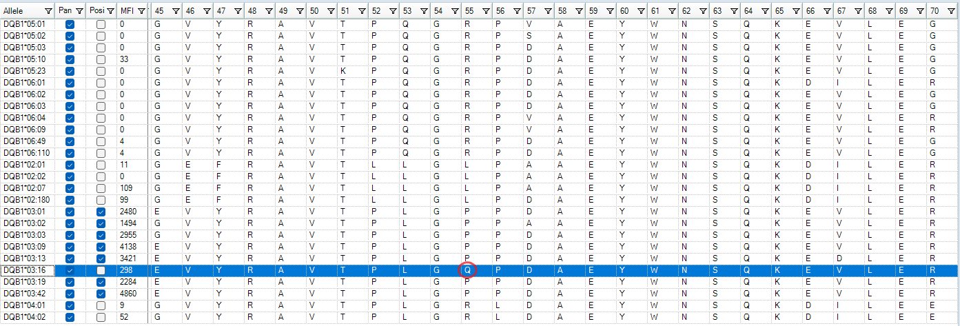
Figure 10: DQB1*03:16 amino acid sequence (HLA Fusion™ Amino Acid module Sequence viewer) highlighting unique glutamine (Q) residue at position 55 as compared to other DQ3 broad group antigens on the panel.
Laboratory workflow
The LABScreen™ Single Antigen HLA Class II ExPlex – Group 2 workflow is unchanged from the standard bead panel. As with the existing HLA Class II ExPlex panel, it can be combined with the standard HLA Class II panel for single-well setup (Figure 11) or run separately and the data later combined in HLA Fusion™ for analysis. Acquisition is using the LABScan 3D™ instrument. This extended HLA-DQ panel benefits from the increased functionality of HLA Fusion™ 4.7.x with regards to use of the Epitope Panel and Amino Acid module.

Figure 11: LABScreen™ Single Antigen HLA Class II ExPlex – Group 2 single-well setup workflow.
Summary
With increasing numbers of patients requiring post-transplant monitoring and/or re-transplantation, the ability to accurately define HLA-antibody profiles is crucial. Current research is highlighting the particular importance of HLA-DQ alloantibodies in both their frequency and impact on graft outcomes. LABScreen™ Single Antigen HLA Class II ExPlex – Group 2 has been developed as an extended HLA-DQ panel providing additional alpha-beta heterodimer diversity and unique polymorphism coverage, improving epitope analysis resolution. Addressing these areas alongside availability of high-resolution HLA typing can add confidence to HLA-DQ alloantibody calls and pre- and post-transplant decision making.
Contact the VH Bio team by filling in the form below for any questions around LABScreen™ Single Antigen HLA Class II ExPlex – Group 2 or for a demonstration of HLA-Fusion™ 4.7.x epitope-based analysis features (e.g. the Epitope Panel and Amino Acid module).
References
- Böhmig GA et al. Antibody-mediated rejection-treatment standard. Nephrol Dial Transplant. 2025; 1;40(8):1615-1627. doi: 10.1093/ndt/gfaf097.
- Tambur A, Kosmoliaptsis V, Claas F. Significance of HLA-DQ in kidney transplantation: time to reevaluate human leukocyte antigen–matching priorities to improve transplant outcomes? An expert review and recommendations. Kidney International. 2021; 100: 1012-1022
- Renaud Snanoudj et al. Epitope load identifies kidney transplant recipients at risk of allosensitization following minimization of immunosuppression. Kidney International. 2019; 95(6): 1471-1485. doi: 10.1016/j.kint.2018.12.029
- Bezstarosti S, Meziyerh S, Reinders MEJ et al. HLA-DQ eplet mismatch load may identify kidney transplant patients eligible for tacrolimus withdrawal without donor-specific antibody formation after mesenchymal stromal cell therapy. HLA. 2023; 102(1): 3-12. doi:10.1111/tan.15008
- Taube, D. et al. De Novo DQ Donor-Specific Antibodies Are Associated With a Significant Risk of Antibody-Mediated Rejection and Transplant Glomerulopathy. Transplantation. 2012; 94(2): 172–177. doi:10.1097/TP.0b013e3182543950
- Meneghini M, Tambur AR. HLA-DQ antibodies in alloimmunity, what makes them different? Curr Opin Organ Transplant. 2023; 1;28(5): 333-339. doi: 10.1097/MOT.0000000000001079
- Ursule-Dufait, C. et al. Preformed Anti-DQ Alpha Donor-specific Antibodies and the Risk of Antibody-mediated Rejection After Kidney Transplantation. Transplantation. 2026; published ahead of print. doi: 10.1097/TP.0000000000005797
- Gupta, V. et al. De novo donor-specific DQA1 antibodies leading to antibody-mediated rejection after renal transplantation. Asian Journal of Transfusion Science. 2025; published ahead of print. doi: 10.4103/ajts.ajts_76_24
- Rene J. Duquesnoy. A Structurally Based Approach to Determine HLA Compatibility at the Humoral Immune Level. Human Immunology. 2006; 67 (11): 847-862. doi:10.1016/j.humimm.2006.08.001.
- Sypek MP, Hughes P. HLA Eplet Mismatches in Kidney Transplantation: More Than Just Adding Things Up. Kidney Int Rep. 2021; 1;6(6):1500-1502. doi: 10.1016/j.ekir.2021.04.027
- Tambur, A. R. et al. (2020). Eplet Mismatch Load and De Novo Occurrence of Donor-Specific Anti-HLA Antibodies, Rejection, and Graft Failure after Kidney Transplantation: An Observational Cohort Study. Journal of the American Society of Nephrology. 2020; 31(9), 2193–2204. doi:10.1681/ASN.2020010019
- Devriese M et al. The Hunt for HLA-DQ Allogeneic Eplets Is Not Over: Four New Ones, Including a Cross-Chain Eplet. HLA. 2025;106(3):e70386. doi: 10.1111/tan.70386.
- Devriese M et al. Characterisation of the HLA-DQ alpha eplet 52SK targeting the DQA1*01 alleles family. HLA. 2024; 104(4):e15700. doi:10.1111/tan.15700. Erratum in: HLA. 2025 Jul;106(1):e70302. doi: 10.1111/tan.70302.
- R. Shih et al. HLA Class I Peptide Polymorphisms Contribute to Class II DQβ0603:DQα0103 Antibody Specificity. Nature Communications. 2024; 15(1): 609. doi: 10.1038/ s4146 7-024-44912-0.
- Tambur, A. R. et al. Eplet Mismatch Load and De Novo Occurrence of Donor-Specific Anti-HLA Antibodies, Rejection, and Graft Failure after Kidney Transplantation: An Observational Cohort Study. Journal of the American Society of Nephrology. 2020; 31(9), 2193–2204. doi.org/10.1681/ASN.2020010019
- Maguire Cet al. Qualitative, rather than quantitative, differences between HLA-DQ alleles affect HLA-DQ immunogenicity in organ transplantation. HLA. 2024; 103(4):e15455. doi:1111/tan.15455
- Van Den Broek, D.A.J., Agrawal, A., Crespo, M. et al. A multicenter kidney transplantation study identifies hierarchy and directionality of HLA-DQ alloimmune responses. Nat Commun. 2026. doi:10.1038/s41467-026-76912-7
Enquire about this article
"*" indicates required fields
