Tissue Slides for Fluorescent Microscope Setup, Calibration, and Performance Validation
OptiSlides Tissue Slides are high-performance fluorescent reference slides designed to support microscope setup, troubleshooting, and system validation across a wide range of light microscopy platforms. Developed by scientists, for scientists, these tissue slides combine rich biological complexity with crisp, multi-channel fluorescence for accurate performance assessment and demonstration of imaging capabilities.
Designed for Real-World Imaging Performance
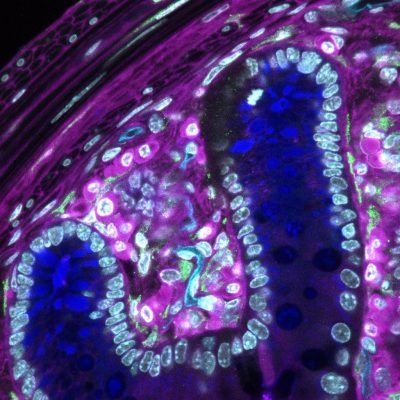
Our standard tissue slide features mouse colon, available in 4-colour, 4-channel and 6-channel configurations (including Near Infrared fluorophores for 735–785 nm laser lines). The sample is stained to highlight:
- DAPI (nuclei, blue)
- PGP9.5 (neurons, green)
- White blood cells (magenta)
- Blood vessels (cyan)
The resulting image, shown here as a maximum intensity projection from a laser scanning confocal microscope (10x objective), captures biologically relevant structures in stunning detail.
6-Channel Near Infrared (NIR) Variants Available
OptiSlides are also available in 6-channel configurations incorporating Near Infrared (NIR) fluorophores, extending performance validation into longer wavelengths for advanced multiplex imaging and spectral separation.
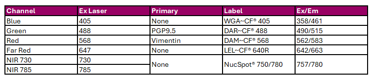
Mouse Colon 6-Channel NIR
Channels and Targets Include:
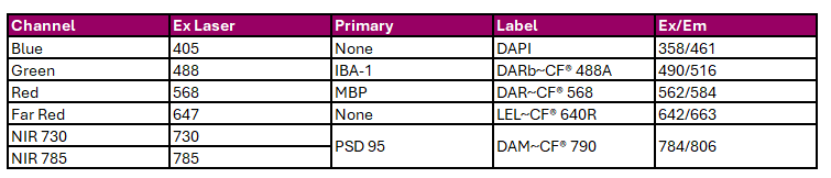
Mouse Brain 6-Channel NIR
Channels and Targets Include:
These variants are designed to support:
- Chromatic integrity assessment across visible and NIR channels
- Depth penetration validation
- Accurate image registration in X, Y, and Z
- Multi-channel alignment and bleed-through evaluation
Validated for use on NIR-enabled fluorescence microscopy systems, including confocal and widefield platforms.
Microscopy Applications
OptiSlides Tissue Slides are compatible with nearly all major light microscopy platforms, including:
- Laser Scanning Confocal
- Spinning Disk Confocal
- Multiphoton
- Widefield and Brightfield
- STED Super-Resolution
- Darkfield and Polarised Light Systems
These slides are ideal for:
- Validating filter sets and camera settings
- Assessing chromatic fidelity and image registration (X, Y, Z planes)
- Demonstrating multi-channel and spectral imaging capabilities
Tissue Thickness Options
- Standard thicknesses: 100 μm and 200 μm
- Ultra-thick samples: Up to 400 μm (available in 2-colour format by request)
- Mounted in VectaShield Plus liquid medium using double-sided adhesive spacers
- Covered with a #1.5 coverslip, sealed with polish or sealant
These thick tissue sections provide an excellent model for testing optical sectioning, depth penetration, and Z-stack acquisition capabilities on advanced imaging systems.
Reusable and Easy to Maintain
- Gently blot immersion oil using Kimwipes® or lens paper
- Clean with 70% ethanol using a cotton-tipped applicator
- Takes just a minute, leaving the coverslip clean and ready for re-imaging
Enquire below to request a quote or more information.
Tissue: Mouse Colon and Brain Prepared Microscope Slides
SKU CODE: 07OPTI-COL-100-VIRTUAL
| PRODUCT | Units |
|---|---|
| Tissue: Mouse Colon, 4 colour, 200 µm07OPTI-COL-200 | Each |
| Tissue: Mouse Brain, 4 colour, 100 µm07OPTI-BRN-100 | Each |
| Tissue: Mouse Brain, 4 colour, 200 µm07OPTI-BRN-200 | Each |
| Tissue: Mouse Brain, 6 colour NIR, 100 µm07OPTI-BRN-100-6 | Each |
| Tissue: Mouse Heart, Unstained for SHG, 100 µm07OPTI-HRT-100-U | Each |
| Tissue: Mouse Heart, Unstained for SHG, 200 µm07OPTI-HRT-200-U | Each |
| Tissue: Mouse Colon, 4 colour, 100 µm07OPTI-COL-100 | Each |
Showing 7 Products
Showing 7 Products
Enquire about this product
"*" indicates required fields