
High-performance PGP 9.5 antibodies for the detection of neurons, neuroendocrine cells, and small nerve fibres in immunohistochemistry (IHC) applications.
Protein Gene Product (PGP 9.5) is an abundant cytoplasmic neuron and neuroendocrine cell-specific protein, recognised as a key marker for neurons and the diffuse neuroendocrine system (DNES). PGP 9.5 appears to belong to a family of ubiquitin–carboxy terminal hydrolase isoenzymes, but while it shows activity towards synthetic ubiquitin esters, the in vivo substrate(s) remain unknown.
| ATTRIBUTE | DETAIL |
|---|---|
| Target | PGP 9.5 / UCHL1 |
| Host | Rabbit |
| Format | Affinity purified |
| Isotype | Rabbit IgG |
| Ractivity | Human, Mouse, Rat |
| Applications | WB, ELISA, paraffin-embedded sections, cryostat sections |
Protein Gene Product (PGP 9.5) is an abundant cytoplasmic neuron and neuroendocrine cell-specific protein, recognised as a key marker for neurons and the diffuse neuroendocrine system (DNES). PGP 9.5 appears to belong to a family of ubiquitin–carboxy terminal hydrolase isoenzymes, but while it shows activity towards synthetic ubiquitin esters, the in vivo substrate(s) remain unknown.
PGP 9.5 antibodies from Cedarlane are meticulously designed to provide superior specificity and performance for immunohistochemistry (IHC) and research applications, including:
- neuropathology
- neuroendocrine studies
- experimental studies of peripheral tissue innervation
- studies of the development and pathology of the DNES
- small fibre neuropathy IHC workflows
PGP 9.5 antibody applications
Antibodies against this protein will stain:
- neuronal cell bodies and axons in the central and peripheral nervous systems
- small nerve fibres in nerve tissue
- neuroendocrine cells in the pituitary, thyroid, pancreas etc.
tumours associated with the diffuse neuroendocrine system
PGP 9.5 antibodies are particularly suitable for detecting small nerve fibres in peripheral tissues. PGP 9.5 immunostaining is widely used in IENFD workflows for the assessment of small nerve fibres in skin biopsy research and small fibre neuropathy studies.
Immunohistochemical analysis of Paraffin-Embedded Sections
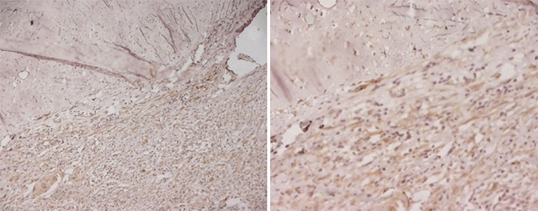
CL7755AP (Anti-Human PGP 9.5) staining (10 µg/ml) of a human cerebral cortex formalin-fixed, paraffin-embedded tissue section; seen at 10x (left) and 20x (right) magnification. Staining on normal brain cortex is negative. High grade tumour (glioma) shows sharp nuclear and cytoplasmic staining in more than 70% of the lesion.
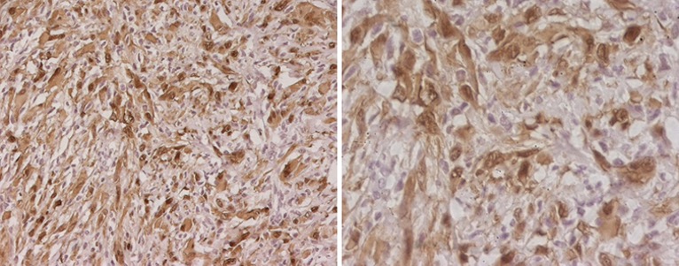
CL7756AP (Anti-Human PGP 9.5) staining (10 µg/ml) of a human cerebral cortex formalin-fixed, paraffin-embedded tissue section; seen at 20x (left) and 40x (right) magnification. Staining on normal brain cortex is negative. High grade tumour (glioma) shows sharp nuclear and cytoplasmic staining in more than 70% of the lesion.
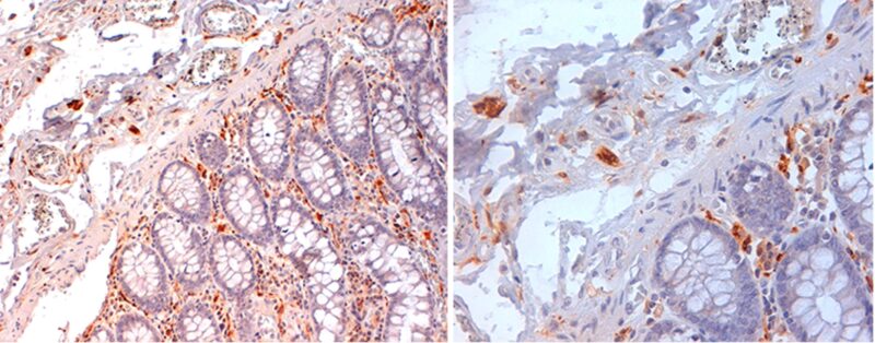
CL7756AP (Anti-Human PGP 9.5) staining (2 µg/ml) of a human colon formalin-fixed, paraffin-embedded tissue section; seen at 20x (left) and 40x (right) magnification. Cytoplasmic/membrane staining of peripheral nerve/ganglion and stromal cells are observed.
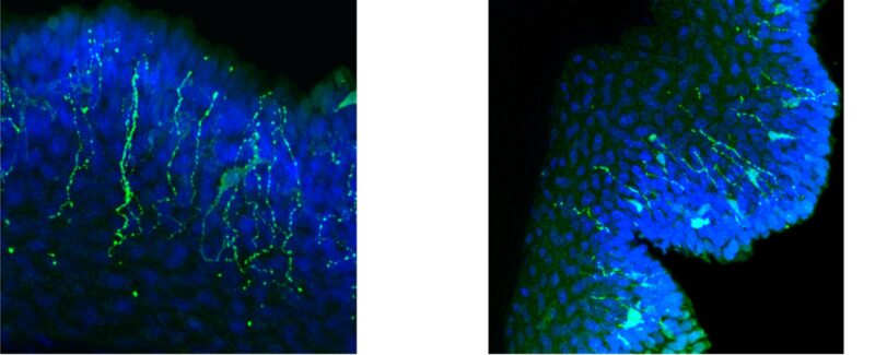
CL7756AP (Anti-PGP 9.5) staining (0.1 µg/ml) of rat epidermis tissue sections isolated from rat paw using thermolysin. The tissue was frozen in M1 embedding matrix and cut on a cryostat to 12 µm. It was fixed in a 0.1 M solution of PBS containing 0.2% (w/v) paraformaldehyde and 0.75% (w/v) picric acid. Sections were imaged using confocal microscopy. Intraepithelial nerve staining of PGP 9.5 is observed (green); tissue was counterstained with DAPI (blue). Image provided by Michael Anderson from Oklahoma State University, Center for Health Sciences. PhD student.
Immunofluorescence analysis
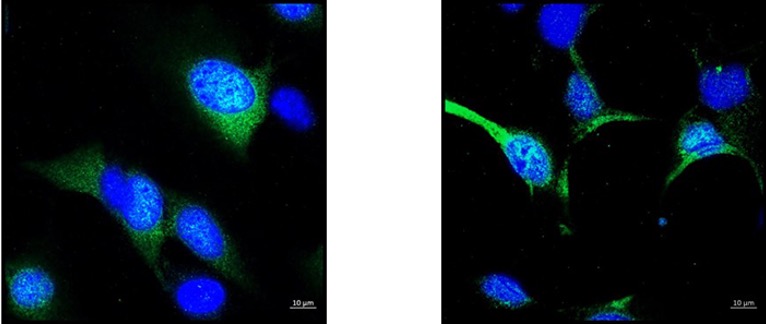
Anti-PGP 9.5 (CL7755AP) staining using goat anti-rabbit Alexa-488 secondary antibody on Adult Mouse Hypothalamus Cell Line mHypoA-2/12 cells (CLU177) imaged under confocal microscope with the 40X oil objective; nuclei were counterstained with DAPI (blue).
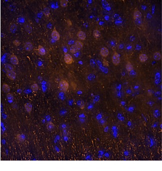
Western Blot Analysis
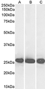
CL7755AP (0.1µg/ml) staining of Human Cerebellum (A), Mouse Brain (B) and Rat Brain (C) lysates (35µg protein in RIPA buffer). Primary incubation was 1 hour. Detected by chemiluminescence.
CL7755AP (0.1µg/ml) staining of Human Cerebellum (A), Mouse Brain (B) and Rat Brain (C) lysates (35µg protein in RIPA buffer). Primary incubation was 1 hour. Detected by chemiluminescence.
Need pricing, availability or technical information? Complete the form below and our team will help you identify the most suitable PGP 9.5 antibody format for your application.
Anti-Human PGP 9.5 Antibodies for IHC, IF and Western Blot
| PRODUCT | Unit Size |
|---|---|
| Anti-Human PGP 9.5 (UCHL1) (199), (affinity purified) (Rabbit IgG)CL7755AP | 100 µg |
| Anti-Human PGP 9.5 (UCHL1) (199), (affinity purified) (Rabbit IgG)CL7755AP-S | 20 µg |
| Anti-Human PGP 9.5 (UCHL1) (184), (affinity purified) (Rabbit IgG)CL7756AP | 100 µg |
| Anti-Human PGP 9.5 (UCHL1) (184), (affinity purified) (Rabbit IgG)CL7756AP-50 | 50 µg |
| Anti-Human PGP 9.5 (UCHL1) (184), (affinity purified) (Rabbit IgG)CL7756AP-S | 20 µg |
Showing 5 Products
Showing 5 Products
Enquire about this product
"*" indicates required fields
