

Fibroblast: DAPI (blue), Peroxisomes (magenta), Paxillin (cyan), Phalloidin (grey)
In scientific research and diagnostics, precision, clarity, and reliability are crucial. The tools you use to conduct experiments and make observations play a pivotal role in determining the accuracy and success of your findings. That’s where Luxidea’s OptiSlides come into play. These advanced fluorescent microscopy slides are specifically designed to test, validate, and showcase high-performance microscopes. Now available exclusively through VH Bio in the UK and Ireland, Luxidea’s OptiSlides have been created by scientists for scientists, ensuring your laboratory’s equipment is always at its best.
Luxidea’s OptiSlides differ from ordinary microscope slides. Prepared with fluorescent markers, they are designed for quick setup, troubleshooting, and demonstrating advanced microscopy systems. Whether you’re using brightfield, widefield, polarised light, confocal, multiphoton, or even STED super-resolution systems, OptiSlides provide the perfect solution to ensure your microscope’s performance. With up to five distinct colours, including spectral options, OptiSlides are essential for scientists working across a wide range of imaging applications.
With tissue thicknesses reaching up to 400 microns, Luxidea’s OptiSlides go beyond conventional three-colour slides, offering a comprehensive solution for testing and calibration in modern laboratories.
A Versatile Lineup for Diverse Research Needs
Luxidea’s OptiSlides lineup features a variety of cell, tissue, and plant slides that replicate real-world laboratory conditions. This ensures accurate testing and validation of microscopes across different biological and environmental samples.
Cell Slides: Customisable, Clear, and Advanced
Luxidea’s cell slides are designed with cellular architecture in mind. Two key options include mouse fibroblast, 4-colour with Paxillin and mouse fibroblast, 4-colour with Vimentin, making them ideal for cytoskeletal and structural studies. These slides also feature near-infrared fluorescence (740Atto) and sub-resolution targets such as peroxisomes (≈150nm), allowing for the highest degree of detail and precision.
These slides replace outdated three-colour models and can be customised to meet specific research needs, ensuring that your microscope’s capabilities are fully validated before analysing critical samples.
Tissue Slides: Replicating Real-World Lab Conditions
For more complex tissue studies, Luxidea’s tissue slides offer unparalleled flexibility. While the colon tissue slide serves as the standard for general testing, Luxidea also offers slides featuring specialised tissues such as liver, lung, lymph node, brain, heart, spleen, testes, and even kidney upon request. These slides are available in up to four colours and thicknesses from 100 to 400 microns, allowing researchers to simulate a wide range of tissue depths for accurate validation.
These tissue slides replace the traditional, thin, three-colour kidney slides, providing a far more detailed solution for testing and calibrating modern microscopes.
Plant Slides: Stone Pine Needle – A Spectral Powerhouse
For plant-based studies, the Stone Pine Needle slide is a unique asset. This slide expresses natural autofluorescence across the spectrum, from ultraviolet (UV) to near-infrared (NIR), making it compatible with nearly any light microscope system. Its multicolour autofluorescence offers distinct features across different channels, and its ability to fluoresce in the far red makes it ideal for fluorescence microscopes equipped with far-red detection.
With thicknesses up to 400 microns, the Stone Pine Needle slide outperforms the traditional convallaria slide, making it ideal for advanced spectral imaging and testing multi-colour registration.
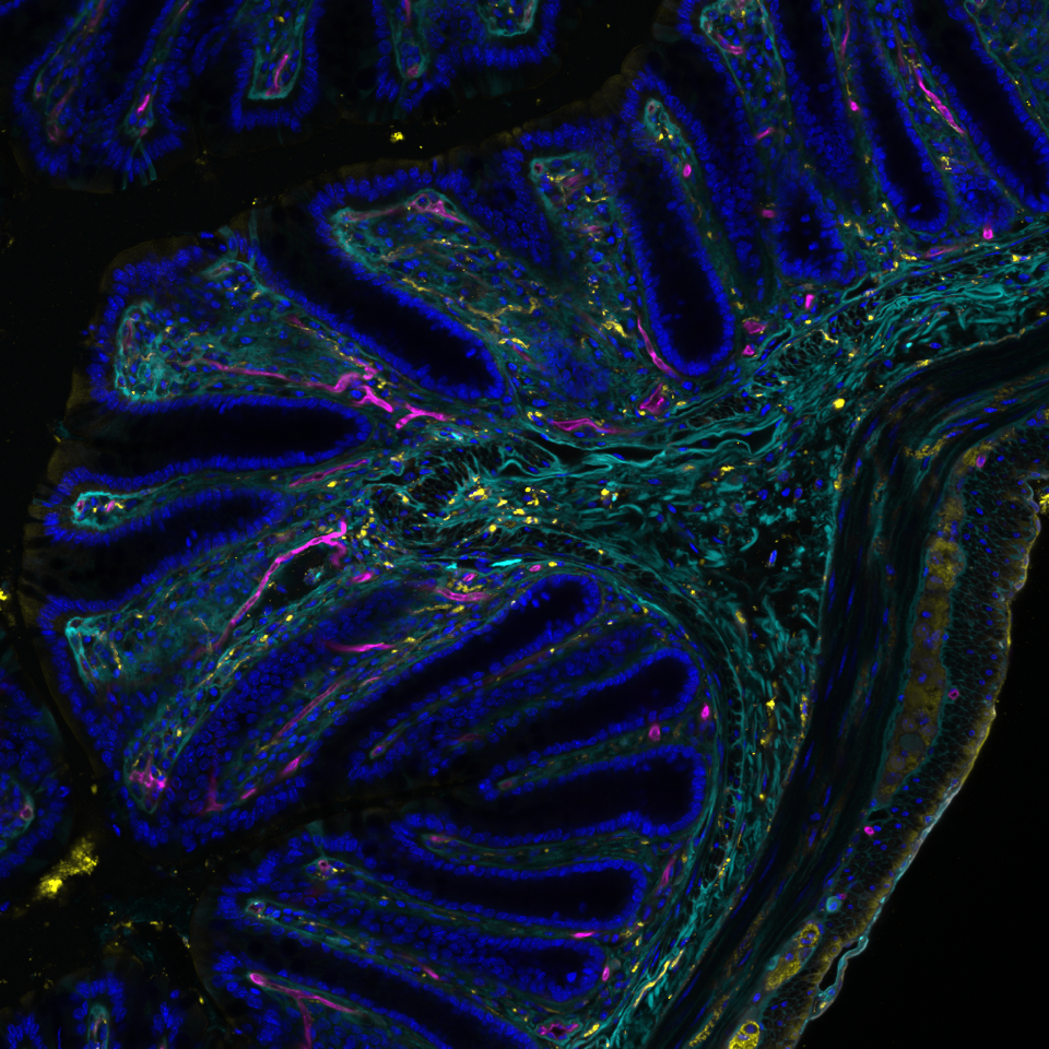
Colon: DAPI (blue), Neurons (yellow), Vimentin (green), Blood Vessels (magenta)
The Value of OptiSlides for Research and Diagnostics
Luxidea’s OptiSlides are crafted to meet the needs of scientists requiring reliable tools for testing, validating, and calibrating their microscopes before conducting critical sample analysis. By ensuring your equipment is performing at its best across a wide range of microscopy techniques, you can focus on what really matters: your research.
In histology and pathology, where the ability to detect small cellular anomalies is critical, OptiSlides ensure your microscope is ready for accurate and detailed analysis. Whether you’re identifying cancerous cells or examining complex tissue structures, OptiSlides offer the validation needed to deliver precise results.
For microbiologists, OptiSlides ensure vivid and distinct colour reproduction, helping to validate the performance of your system before classifying microorganisms or conducting detailed bacterial analysis.
Cytologists, working with fragile cell samples, benefit from the exceptional clarity of Luxidea’s cell slides, ensuring that microscopes are functioning properly before examining delicate samples. This allows for the detection of subtle cellular abnormalities with greater precision.
In fluorescence microscopy, background interference can severely impact results. Luxidea’s OptiSlides are specifically designed to minimise interference, ensuring that fluorescent signals remain bright and distinguishable. This is especially vital in immunofluorescence studies, where detecting fluorescent markers with pinpoint accuracy is essential.
OptiSlides: A Reliable and Versatile Solution
By offering enhanced optical clarity, flexibility, and performance, these slides allow scientists to confidently test, validate, and calibrate their microscopes across diverse applications—whether in cell, tissue, or plant research. Luxidea’s OptiSlides provide the precision and dependability needed to produce accurate and reliable results in every experiment.
As the exclusive distributor of Luxidea’s OptiSlides in the UK and Ireland, VH Bio is proud to bring this cutting-edge product to your laboratory.
Contact us by filling in the form below for detailed specifications on Luxidea’s high-performance fluorescent microscopy slides, and discover how they can help optimise your laboratory’s performance.
Learn more:
Standard microscope slides vs specialist imaging slides: when does your application need more?
Enquire about this article
"*" indicates required fields